Jasmeet Dhaliwal
Honours Integrated Science (B.Sc.), Class of 2024, McMaster University
dhalij34@mcmaster.ca
Stress is a term used to describe emotionally and physically demanding circumstances. In common jargon, “good stress” pertains to short experiences and leaving a feeling of accomplishment, whereas “bad stress” refers to experiences that lack a degree of stability and are frequently prolonged or emotionally and physically exhausting. The stress system, which combines a wide variety of brain regions that, together, are capable of detecting events and interpreting them as genuine or prospective dangers, is known to construct and initiate the reaction to stressful stimuli. Different stresses activate specific neural networks, necessitating fine-tuned functional neuroanatomical interpretation.1 This quick activation of the Sympathetic-Adreno-Medullar (SAM) and Hypothalamus-Pituitary-Adrenal (HPA) axes, the two crucial components engaged in stress response, may occur from the integration of signals from the stressor directly.1 The stress response’s intricacy is not limited to neuroanatomy or SAM and HPA axis mediators. However, it varies depending on the length of stressor exposure and its short-term and long-term repercussions. This comprehensive review will explore the extensive neuroanatomy involving stress domains and the neuro activity for physical and psychological stressors.
A stressor’s perception is the initial stage in the stress response. When a scenario is perceived as dangerous, the brain activates various neural circuits to maintain physiological integrity even under extreme circumstances. However, multiple networks must be engaged to identify different sorts of stresses. Different neural networks and cellular activity are activated in response to psychological and physical stimuli (Figure 1), producing various ‘footprints’ in the brain.2 Physical stressors are stimuli that cause genuine physiological problems, such as bleeding or infection, and overburden the body. On the other hand, psychological stressors are social and physical environmental situations that pose a threat to an individual’s current state and are regarded as anticipatory.3 Unpleasant external stimuli, predator-related signals, and the inability to fulfil internal urges are a few examples of this stressor. As a result, distinct circuitries in the brain handle physical and psychological stimuli, which may overlap in some cases. Nonetheless, the stress system will be triggered in a synchronized approach regardless of how the stressor is processed.
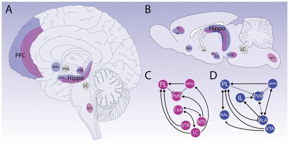
Figure 1: Physical (pink) and psychological (blue) stressors are processed by primarily neuroanatomical substrates, as depicted in this diagram. The upper panels indicate how numerous structures are involved in brain processing for distinct forms of stressor detection and situation appraisal (A, B, respectively). The bottom panels show how physical and psychological pressures necessitate the activation of several networks (C, D, respectively). Both stressors activate different brain regions, subsequently causing a cascade of neurobiological effects. Though these pathways are separate, they do overlap in certain areas such as the prelimbic area (PL) and paraventricular nucleus of the hypothalamus (PVN) (3).
Physical stresses are handled primarily by the brainstem and hypothalamus areas and typically necessitate an early systemic response. Two major systems are triggered when exposed to stressors: first the SAM and then the HPA.3 The neurological system reacts quickly when activating the SAM network, which concerns the adrenal medulla in the autonomic nervous system (ANS) and causes the release of catecholamines (i.e. adrenaline and noradrenaline).4 This triggers an increase in heart rate (HR) and blood pressure (BP), allowing for increased physical activity. Subsequently, the HPA axis is slowly activated, which concerns the adrenal cortex and secretes corticotropin-releasing hormone (CRH) in the sympathetic nervous system (SNS).5,6 Since CRH helps to mobilize the body’s energy through the use of glucose, providing energy to cope with the stressor, however, this hormonal release also contributes to negative effects such as reduced immune function due to the anti-inflammatory effects of CRH.7,8 Both systems are often co-activated and integral to response during a stressful situation for the body to prepare for the threat and then return to a stable state once the threat has passed.8 Therefore, when a real stressor is identified or interpreted by the brainstem through signals, the two major networks are activated alongside other structures for triggering the stress response.
Physically demanding stressors are more likely to elicit autonomic stress, whereas psychologically unpredictable and threatening stress produce physical and behavioural stress. The prefrontal cortex (PFC) is essential for understanding appropriate reactions to environmental changes and facilitating behavioural plasticity.9 However, since distinct anatomic subdivisions perform diverse functions, the role of the prefrontal cortex (PFC) in stress response is complex. The amygdala, an essential region related to emotional processing, contains substantial connections from the PFC.10 Essentially, the whole amygdala complex appears to stimulate corticosteroid production and release. The basolateral nucleus (BLA), the central nucleus of the amygdala (CeA), and the medial nucleus (MeA) encompass the amygdala complex.4 The BLA is a critical player in processing psychological stresses, and anticipatory stressors predominantly activate it.7 The CeA has also been linked to physiological reactions to frightening stimuli, stressful stimuli, and even drug-related stimuli.10 Both structures have a large population of projecting neurons with CRH receptors, which regulate pituitary adrenocorticotropic hormone (ACTH) secretion and mediate behavioural and autonomic responses to stress by interacting with type 1 plasma membrane receptors (CRHR1) in pituitary corticotropes and the brain.11 Thus, a stressful situation, whether environmental or emotional, can activate the PFC and trigger a cascade of stress hormones to be released.
The physiologic reactions of the body to stress are essential in the therapeutic context for a variety of reasons, including the care of healthy and hypo adrenal surgery patients and determining how patients’ lifestyle changes may affect the body’s stress response.12,13 This review explains that various components, such as hormones, receptors, and neurological structures, are involved in the body’s reaction to stress. In order to give an appropriate or optimum treatment approach or intervention for individual patients, it is critical for the physician delivering stress therapy to grasp the relationship between physical and psychological stressors.
- Dayas CV, Buller KM, Crane JW, Xu Y, Day TA. Stressor categorization: acute physical and psychological stressors elicit distinctive recruitment patterns in the amygdala and in medullary noradrenergic cell groups. Eur J Neurosci. 2001;14(7):1143–52.
- Godoy LD, Rossignoli MT, Delfino-Pereira P, Garcia-Cairasco N, de Lima Umeoka EH. A Comprehensive Overview on Stress Neurobiology: Basic Concepts and Clinical Implications. Front Behav Neurosci. 2018 [cites 2022 Feb 25]; Available from: https://www.frontiersin.org/article/10.3389/fnbeh.2018.00127.
- Glavin GB. Stress and brain noradrenaline: a review. Neurosci Biobehav Rev. 1985;9(2):233–43.
- Ulrich-Lai YM, Herman JP. Neural Regulation of Endocrine and Autonomic Stress Responses. Nat Rev Neurosci. 2009;10(6):397–409.
- Seki K, Yoshida S, Jaiswal MK. Molecular mechanism of noradrenaline during the stress-induced major depressive disorder. Neural Regen Res. 2018;13(7):1159–69.
- Janak PH, Tye KM. From circuits to behaviour in the amygdala. Nature. 2015;517(7534):284–92.
- Wang S, Liu X, Shi W, Qi Q, Zhang G, Li Y, et al. Mechanism of Chronic Stress-Induced Glutamatergic Neuronal Damage in the Basolateral Amygdaloid Nucleus. Anal Cell Pathol. 2021 [cited 2022 Feb 25]; Available from: https://www.ncbi.nlm.nih.gov/pmc/articles/PMC8632434//
- Smith SM, Vale WW. The role of the hypothalamic-pituitary-adrenal axis in neuroendocrine responses to stress. Dialogues Clin Neurosci. 2006;8(4):383–95.
- Jie F, Yin G, Yang W, Yang M, Gao S, Lv J, et al. Stress in Regulation of GABA Amygdala System and Relevance to Neuropsychiatric Diseases. Front Neurosci [Internet]. 2018 [cited 2022 Feb 25]; Available from: https://www.frontiersin.org/article/10.3389/fnins.2018.00562
- Roozendaal B, Brunson KL, Holloway BL, McGaugh JL, Baram TZ. Involvement of stress-released corticotropin-releasing hormone in the basolateral amygdala in regulating memory consolidation. Proc Natl Acad Sci. 2002;99(21):13908–13.
- Kolb B, Mychasiuk R, Muhammad A, Li Y, Frost DO, Gibb R. Experience and the developing prefrontal cortex. Proc Natl Acad Sci U S A. 2012;109:17186–93.
- Thau L, Gandhi J, Sharma S. Physiology, Cortisol. In: StatPearls [Internet]. Treasure Island (FL): StatPearls Publishing; 2022 [cited 2022 Feb 25]; Available from: http://www.ncbi.nlm.nih.gov/books/NBK538239/.
- Wang S, Shi W, Zhang G, Zhang X, Ma C, Zhao K, et al. Endoplasmic Reticulum Stress-Mediated Basolateral Amygdala GABAergic Neuron Injury Is Associated With Stress-Induced Mental Disorders in Rats. Front Cell Neurosci. 2019 [cited 2022 Feb 25]; Available from: https://www.frontiersin.org/article/10.3389/fncel.2019.00511.
